Lateral Cephalometry X Ray
Lateral cephalometry is a highly specialized dental and orthodontic imaging technique that captures a complete side-view image of the skull, jaw, and facial bones. Unlike regular dental X-rays that focus mainly on the teeth, lateral cephalometry provides a broader perspective, allowing orthodontists, oral surgeons, and dentists to assess the relationships between different facial structures. This type of imaging is essential for planning orthodontic treatments such as braces or aligners, preparing for corrective jaw surgeries, monitoring facial growth in children, and even diagnosing airway-related conditions like obstructive sleep apnea. The image provides accurate angular and linear measurements that guide precise treatment decisions.
At our Lateral Cephalometry X Ray Centre in Pallavaram, we use advanced digital radiography technology to ensure you receive clear, high-resolution images with minimal radiation exposure. Our machines are designed to capture the exact positioning of teeth, jaws, and facial bones, enabling dental professionals to create personalized treatment plans. The process is quick, painless, and non-invasive, making it suitable for patients of all ages. Whether you are undergoing orthodontic assessment, surgical planning, or follow-up imaging, our centre is equipped to deliver accurate results with the highest standards of safety and comfort.
What is Lateral Cephalometry?
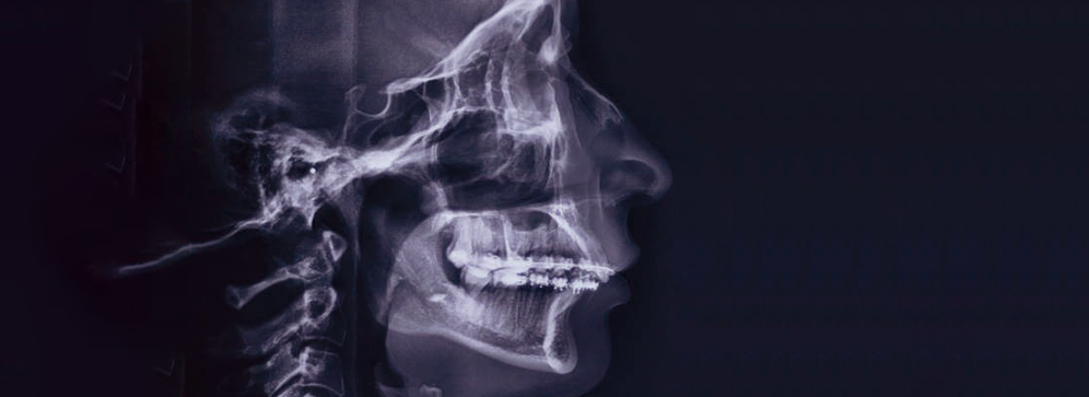
Lateral cephalometry is a diagnostic X-ray that captures a side profile of the head, showing the bones and facial contours in precise detail. It allows dentists and orthodontists to evaluate how the jaw, teeth, and skull relate to one another in terms of position, proportion, and alignment. This information is crucial when deciding whether a patient needs orthodontic appliances, surgery, or other corrective treatments. It is also invaluable in tracking changes over time, such as jaw growth in children or treatment progress in orthodontic cases.
The lateral cephalometric image is more than just a picture; it is a tool for measurement and analysis. Orthodontists can use specialized software to analyze the image, measure bone angles, assess airway size, and predict how treatments will affect facial appearance. Because of its ability to show both hard and soft tissues, it provides a holistic view of the patient’s craniofacial structure. This makes it a key diagnostic tool in modern orthodontics and maxillofacial surgery.
- Detailed side-view X-ray of the head and face
- Shows bone and soft tissue structures clearly
- Essential for orthodontic and surgical planning
- Useful in airway and breathing assessments
- Helps track facial growth and treatment progress
What is Lateral Cephalometry?
Lateral cephalometry is a diagnostic X-ray that captures a side profile of the head, showing the bones and facial contours in precise detail. It allows dentists and orthodontists to evaluate how the jaw, teeth, and skull relate to one another in terms of position, proportion, and alignment. This information is crucial when deciding whether a patient needs orthodontic appliances, surgery, or other corrective treatments. It is also invaluable in tracking changes over time, such as jaw growth in children or treatment progress in orthodontic cases.
The lateral cephalometric image is more than just a picture; it is a tool for measurement and analysis. Orthodontists can use specialized software to analyze the image, measure bone angles, assess airway size, and predict how treatments will affect facial appearance. Because of its ability to show both hard and soft tissues, it provides a holistic view of the patient’s craniofacial structure. This makes it a key diagnostic tool in modern orthodontics and maxillofacial surgery.
- Detailed side-view X-ray of the head and face
- Shows bone and soft tissue structures clearly
- Essential for orthodontic and surgical planning
- Useful in airway and breathing assessments
- Helps track facial growth and treatment progress
How the Procedure Works
Getting a lateral cephalometry X-ray is simple, quick, and comfortable. The patient is positioned in a cephalostat, a device that keeps the head stable during the imaging process to ensure accuracy. The technician will adjust the height and position to align the head with the X-ray beam, often using light markers for guidance. A small plastic bite piece may be used to help maintain jaw alignment during the scan.
Once the patient is correctly positioned, the machine captures the X-ray in just a few seconds. The resulting digital image is processed immediately and can be reviewed by the technician and dentist right away. There’s no need for film development, and the results can be securely shared with your orthodontist or oral surgeon. The entire visit, including preparation, positioning, and scanning, usually takes less than 15 minutes, making it highly convenient for patients with busy schedules.
- Quick and comfortable process
- Head held steady using a cephalostat
- Digital images ready in seconds
- No special preparation required
- Minimal disruption to your day
Advantages of Digital Cephalometry
Digital lateral cephalometry offers significant benefits over older film-based systems. The most obvious advantage is the clarity and resolution of the images. High-definition digital images allow for zooming, rotating, and enhancing specific areas without losing quality. This level of detail enables more accurate diagnoses and treatment planning, which directly impacts the success of orthodontic and surgical outcomes.
Another major benefit is reduced radiation exposure. Modern digital systems use far less radiation compared to traditional X-ray film, making them safer for patients. Additionally, digital images can be stored electronically, eliminating the risk of losing or damaging physical films. They can be shared instantly via secure channels with other dental specialists, ensuring seamless collaboration between healthcare providers. This convenience speeds up decision-making and allows for better-coordinated care.
- Clear, high-resolution imaging for precise diagnosis
- Reduced radiation exposure for patient safety
- Digital storage and instant file sharing
- Eco-friendly process without chemical film development
- Better treatment outcomes due to enhanced image clarity
Uses of Lateral Cephalometry in Dentistry
Lateral cephalometry plays a vital role in orthodontics, where accurate measurements are necessary for planning and monitoring treatment with braces or clear aligners. The scan helps determine the position of the upper and lower jaws, the angulation of the teeth, and how these relate to facial symmetry. This information allows orthodontists to create treatment plans that achieve not only functional but also aesthetic results.
In oral and maxillofacial surgery, lateral cephalometry is used to plan corrective jaw surgeries and evaluate post-operative outcomes. It is also valuable in diagnosing and managing sleep apnea by assessing airway space. Pediatric dentists use it to track craniofacial growth patterns in children, which can help identify and address orthodontic issues early, potentially reducing the need for more complex treatments later.
- Orthodontic treatment planning for braces and aligners
- Surgical planning and post-surgery evaluation
- Airway analysis for sleep disorder diagnosis
- Monitoring jaw growth in children
- Improving facial aesthetics through treatment
Safety Considerations
Safety is a priority when it comes to any X-ray procedure. Modern lateral cephalometry systems are designed to use the lowest possible radiation dose while still producing high-quality images. This ensures that patients, including children, can undergo the scan without unnecessary exposure. The risk from this level of radiation is minimal, especially when compared to the valuable diagnostic information it provides.
Patients who are pregnant or think they might be pregnant should inform the technician before the scan. Additional protective measures, such as lead aprons and thyroid shields, can be used to further reduce exposure. Our technicians follow strict radiation safety guidelines and maintain equipment regularly to ensure safe operation at all times.
- Extremely low radiation exposure
- Safe for adults and children
- Special precautions for pregnant patients
- Use of protective shielding when necessary
- Compliance with international safety standards
Preparing for the Scan
Preparing for a lateral cephalometry scan is simple and stress-free. There are no dietary restrictions, fasting requirements, or special instructions to follow beforehand. The only request is to remove any metallic objects from the head and neck area, such as earrings, necklaces, eyeglasses, or hairpins, as these can distort the image.
During the scan, the technician will provide instructions to ensure proper positioning. Patients should follow these instructions carefully and remain still during the image capture. Staying motionless ensures the resulting X-ray is sharp and clear, eliminating the need for repeat scans. The entire process is comfortable, making it suitable for children and adults alike.
- No fasting or special preparation needed
- Remove metal accessories for accurate results
- Follow technician’s positioning instructions
- Remain still during the scan to avoid blurring
- Quick, easy, and comfortable procedure
Why Choose Our Lateral Cephalometry X Ray Centre in Pallavaram
Our centre is trusted by leading orthodontists, dentists, and surgeons in the Pallavaram area because we combine advanced technology with professional expertise. We understand that accurate imaging is the foundation of effective treatment, so we prioritize precision, clarity, and safety in every scan we perform.
We also pride ourselves on providing a patient-friendly experience. From short wait times to convenient scheduling and immediate results, we ensure that your visit is smooth and efficient. Our central location in Pallavaram makes us easily accessible, and our competitive pricing means you receive top-quality imaging without overpaying.
- Advanced digital radiography systems
- Skilled, experienced radiology technicians
- Immediate image availability
- Patient-focused service and comfort
- Convenient Pallavaram location
Your Smile’s Precision, Our Commitment — at Insight Diagnostics and Labs, we believe that every detail matters when it comes to your dental health and orthodontic planning. Our Lateral Cephalometry X-Ray Centre in Pallavaram is equipped with modern imaging technology and operated by experienced radiology professionals who understand the importance of accuracy in orthodontic assessments. We go beyond simply capturing an image — we ensure every scan is clear, precise, and tailored to the needs of your dentist or orthodontist, helping them create a treatment plan that’s just right for you.
We are committed to delivering a comfortable, safe, and patient-friendly experience for every visitor. Whether you’re visiting us for an orthodontic evaluation, jaw alignment study, or pre-treatment documentation, our team ensures that you feel informed and cared for at every stage. With quick turnaround times, affordable rates, and unwavering quality standards, we make advanced dental imaging accessible to everyone in Pallavaram. Trust us to be your partner in achieving the healthy, confident smile you deserve — because at Insight Diagnostics, your care always comes first.

